Research Highlights
NCMIR researcher wins second and eighth place in the Nikon International Small World Competition.
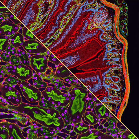
October 2005
Nikon Corp has announced the winners of the 2005 Nikon International Small World Competition. Local NCMIR scientist Tom Deerinck garnered a second and an eighth place in the prestigious competition with his images of tissues stained with fluorescent quantum dots. According to Nikon: “The Nikon Small World competition recognizes the fusion of science and art as captured through the lens of the optical microscope. This year's competition has been fueled by a record number of entries submitted by professional and amateur microscopists from around the globe. These photographers blend their technical and artistic talents to produce images that capture the imagination and spark scientific curiosity.”
Judges chosen by Nikon for the competition include: Jennifer Waters, PhD, Microscopy Director at Harvard Medical Center; Emily Harrison, Photography Editor of Scientific American Magazine; Todd James, Illustrations Editor of National Geographic Magazine; Alexey L. Khodjakov, PhD, Research Scientist at the Wadsworth Center; and, guest consultant Michael Davidson, Senior Research Engineer, National High Magnetic Field Laboratory at Florida State University. Nikon recognized the Small World winners at an event on October 6, 2005 in New York City. The winning images will be exhibited during the course of 2006 in several formats including an annual color calendar, a national museum tour and an electronic gallery. For more information see: http://www.nikonsmallworld.com