Research Highlights
NCMIR’s Electron Tomography Resource Reveals Novel Mitochondrial Anchoring Scaffolds and Cristae Structured for High-rate Metabolism
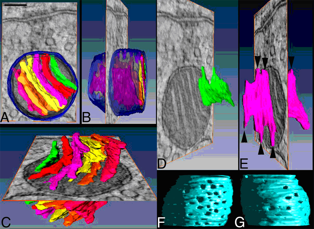
Caption: Figure. A. MAC mitochondria have cristae that fill their interior. A top-view of the surface-rendered volume is superimposed over a roughly central slice through the reconstruction shown for perspective. The outer mitochondrial membrane (OMM) is shown in translucent blue and each crista in a different color. B. A side-view of the surface-rendered volume. C. MAC mitochondria have only lamellar cristae that are not interconnected. D, E. Examples from two cristae are shown. The lamellar cristae have “fingers” that connect to the periphery via circular openings and are termed crista junctions (arrowheads). F, G. Views of inner mitochondrial membrane with OMM removed. Crista junctions have a polar distribution within MAC mitochondria. There are more junctions along the presynaptic face (viewed from presynaptic membrane in panel F) than along the opposite face (viewed from interior of terminal in panel G). The openings are visualized using right-lighting of a side view. Panel A scale bar = 100 nm.
January 2010
January 2010 – Electron microscope tomography was used at the NCMIR to aid Prof. George Spirou (West Virginia University) in his investigation of the mitochondria-associated adherens complex (MAC) in the auditory brain stem and resulted in a publication in the J. Neuroscience. In nerve terminals, mitochondria must be appropriately positioned to regulate microdomains of Ca2+ concentration and metabolic demand, but structures that anchor them in place have not been described. By applying high-resolution electron tomography (ET) to the study of a central terminal, the calyx of Held, researchers at the NCMIR revealed an elaborate cytoskeletal superstructure that connects a subset of mitochondria to the presynaptic membrane near active zones. This cytoskeletal network extended laterally and was well integrated into the nerve terminal cytoskeleton, which included filamentous linkages among synaptic vesicles. ET revealed novel features of inner membrane for these mitochondria (figure). Cristae structure was polarized in that crista junctions, circular openings of the inner membrane under the outer membrane, were aligned with the cytoskeletal superstructure and occurred at higher density in mitochondrial membrane facing the presynaptic membrane. These characteristics represent the first instance where a subcomponent of an organelle is shown to have a specific orientation relative to the polarized structure of a cell. The ratio of cristae to outer membrane surface area is large in these mitochondria relative to other tissues, indicating a high metabolic capacity. These observations suggest general principles for cytoskeletal anchoring of mitochondria in all tissues, reveal potential routes for non-synaptic communication between pre and postsynaptic partners utilizing this novel cytoskeletal framework, and indicate that cristae structure can be specialized for particular functions within cellular microdomains.
Full text: http://www.ncbi.nlm.nih.gov/pubmed/20089910?dopt=Abstract
Related Publication
Perkins, G.A., Tjong, J., Brown, J.M., Scott, R.T., Poquiz, P.H., Ellisman M.H. and Spirou, G.A. (2010) The micro-architecture of mitochondria at active zones: Electron tomography reveals novel anchoring scaffolds and cristae structured for high rate metabolism. J. Neurosci., 30:1015-1026.