Research Highlights
NCMIR Develops New Technique to Improve Resolution of SBEM Images
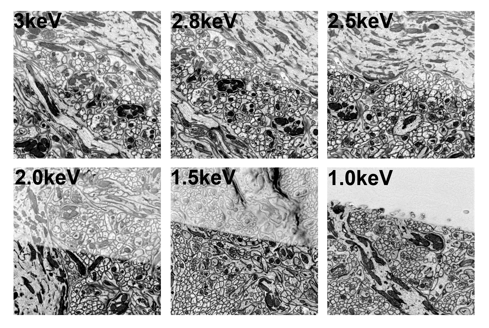
Electron-penetration depth as a function of probe-beam landing energy, modulated via a decelerating biasing potential. A 30 nm thick blank plastic section was placed on top of a cerebellum block sample. Images were then collected using a FEI CBS detector with the central ring turned off. The 30 nm section runs diagonally across the middle of the image. Images are inverted so white shows no backscattering signal. All images were acquired with 3-keV column electron energy and deceleration appropriate to achieve the listed electron-landing energies. (A) 3-keV landing energy. (B) 2.8-keV landing energy. (C) 2.5-keV landing energy. (D) 2-keV landing energy. (E) 1.5-keV landing energy. (F) 1-keV landing energy. At a landing energy between 1.5 keV and 1 keV, the electron penetration depth drops below 30 nm.
15-09-2016 La Jolla, CA
Three-dimensional imaging across spatial scales is an important tool to help researchers understand biological structure. In this context, serial block-face scanning electron microscopy (SBEM) has become a key technology to elucidate structure in the moderate lateral and axial resolution range (10 and 40 nm, respectively) over wide fields of view. As such, it has proven useful for neuron tracking.
With volume imaging, researchers not only wish to increase the size and extent of tissue volumes imaged, they also seek ways to improve resolution and the signal-to-noise ratio. This article describes one new approach to do that.
But first some background on SBEM. An SBEM platform uses a microtome to slice ~30-80nm-thick sections from the sample block. After each slice is removed, the electron-beam scans the newly exposed blockface at a defined accelerating voltage, and backscattered electrons (BSEs) are then collected by a detector to create an image of the newly exposed blockface. This process continues, section by section, until the complete tissue volume is imaged.
Computationally integrating all the images together in the order they were created produces a 3D volume that can then be analyzed to understand more about the internal structure of the sample. The beauty of this approach is that the digital sample continues to “live”: It can be sliced and diced repeatedly by multiple researchers working on their own projects, even well into the future as new scientific questions emerge.
In SBEM, scattering of electrons in the sample is governed principally by the landing energy of the probe-beam electrons and composition of the sample. Limits to resolution include the interaction volume of the electrons into the sample, sample charging, the number and profile of the BSEs (i.e., how far the BSEs emerge from the beam impact point and the fraction of the beam energy they have), and the thickness of the ultramicrotome cuts.
One way to improve image resolution is to lower the energy of the probe-beam electrons. At lower energies (e.g., 2.0 keV or lower), the volume of the sample that the electrons interact with is much smaller, so surface detail should be more highly resolved. However, at these lower energies, the BSE yield and detector sensitivity are both sub-optimal, thus negating the anticipated gain in resolution.
But a team of researchers at NCMIR, led by James C. Bouwer, found a way to address this challenge. They applied a negative voltage to a SBEM stage to create a tunable electric field at the surface of the sample, which was used to decrease the electron landing energy and alter the trajectory of the BSEs. This approach reduced electron penetration in the sample to less than 30 nm, accompanied by re-acceleration of the BSEs toward the detector, leading to a large increase in the signal that the detector recorded.
Results showed signal level increases of up to 20x and 6x or greater at 1- and 1.5-keV landing energy, respectively, for decelerated probe-beam electrons versus those in conventional SBEM. Moreover, the increase in the signal-to-noise ratio provided by the acceleration of the BSEs reduced the needed dose per unit area and enabled significantly faster scan rates at higher magnifications.
By tuning the electric field, the researchers could also manipulate the trajectories of the BSEs and secondary electrons (SEs), enabling spatial discrimination between them. This discrimination enabled separating out the SEs, which generally provide unsatisfactory contrast, from the BSEs recorded by the detector and key to creation of the image. The researchers blocked SEs using an FEI concentric backscatter detector and a simple SE-blocking aperture in a monolithic detector.
These approaches make probe-beam deceleration a feasible approach to achieve limited electron sample penetration with sufficient imaging stability to produce large volumes of more highly resolved data recorded by the detector.
Funding Source: NIH National Institute of General Medical Sciences award P41GM103412 to Mark H. Ellisman to operate NCMIR. The authors thank Gatan, FEI Company, and Carl Zeiss for the loan of SBEM equipment used in this study.
Relevant Publication: Bouwer, James C., Thomas J. Deerinck, Eric Bushong, Vadim Astakhov, Ranjan Ramachandra, Steven T. Peltier, and Mark H. Ellisman. "Deceleration of probe beam by stage bias potential improves resolution of serial block-face scanning electron microscopic images." Advanced Structural and Chemical Imaging 2, No. 1 (2016): 11.