Research Highlights
A New Framework for the Formation and Development of Learning-related Dendritic Spines
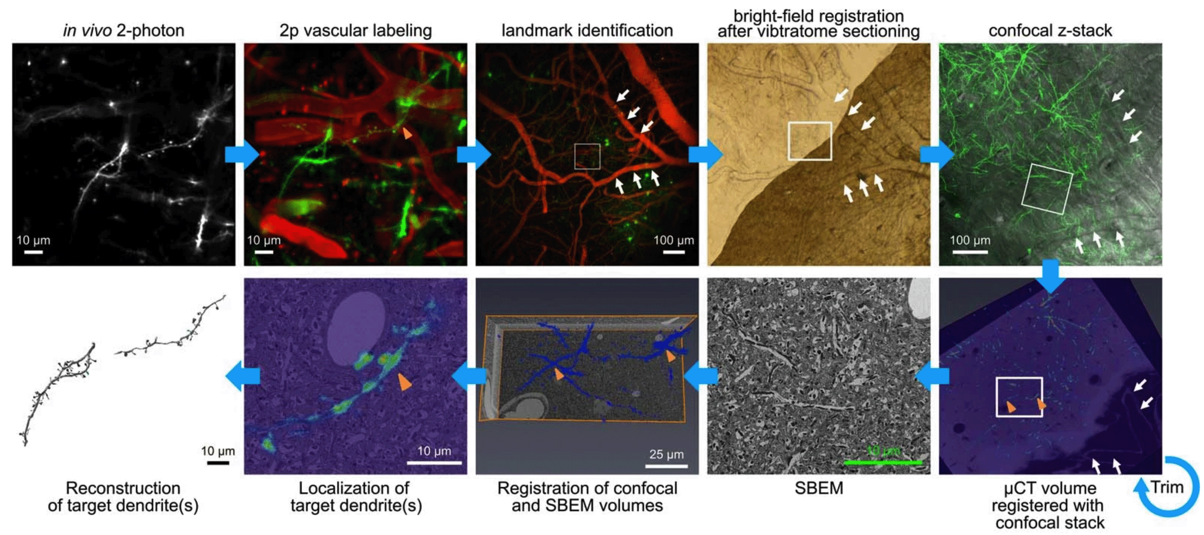
Workflow of correlated light and electron microscopy (CLEM) method to identify the in vivo-imaged dendrites for EM.
02-06-2022 La Jolla, CA
Learning is associated with the formation of new synaptic connections between neurons in the brain. This is thought to shift how neurons behave based on the new information that they receive. In truth, however, very little is known about the function of new synapses during learning, and the principles used by the brain to efficiently make useful new connections are poorly understood.
In recent work published in Nature Neuroscience, Hedrick et al. used 2-photon microscopy to visualize individual synapses in actively learning mice and tracked these structures over days of repeated learning, allowing them to identify new synapses that formed over the process. By using a fluorescent biosensor that reflects synaptic activity, they also measured the functional behavior of synapses during learning, allowing them to detail how the information carried by new synapses compared to the rest of the population.
Remarkably, they observed that new synapses were typically grouped together in space with other synapses that showed similar synaptic activity (i.e. that carried similar information), suggesting that the process of forming new connections also involves keeping them organized according to their function. What results from this organization are ‘clusters’ of synapses that are close together and whose activity is synchronized in time. This synchronized activity appeared to signal components of the learned behavior, suggesting that new synaptic connections tend to support behavior primarily by cooperating with other synapses.
To understand more about how this organization arises, and what its specific consequences might be, the researchers performed a technique called correlated light- and electron microscopy (CLEM) to study the tiny cellular details surrounding synapses that they tracked in the living brain. Using this technique, they found evidence for a series of basic cellular functions that ensure the organization of new synapses. Specifically, there were hints that the strengthening of some synapses during early stages of learning simultaneously gives rise to the nearby outgrowth of “candidate” new synapses. While most candidates likely fail to ever make a synapse, a portion of them successfully found connections, but critically, only those that were synchronized with nearby synapses survived. Thus, the strengthening of specific information during early learning gives rise to a cascade of events that amplify the target information through the addition of synchronized new synapses nearby. While this model would seem to favor new synapses simply duplicating the early-phase target information, the authors instead found that new synapses tended to make connections that were previously not represented in the cluster.
These data suggest that new synaptic connections guide learning by incorporating distinct inputs carrying similar information into synchronized synaptic clusters. As a whole, these findings provide a plausible biological mechanism by which neurons can organize new information according to their existing structure, thereby making efficient use of small changes and likely granting the brain flexibility while remaining robust to confusion.
The original manuscript can be found here (DOI: 10.1038/s41593-022-01086-6).
Links to recent press:
https://medicalxpress.com/news/2022-06-framework-formation-learning-related-dendritic-spines.html